Histopathology-Cytology Equipment
About
An accurate diagnosis relies on the correct processing of tissue, completed with the maximum care. The proper collection and preparation of samples, according to International Guidelines and Protocols, is critical to ensure the integrity of the tissue and proteins of interest, and to enable the detection of antibodies.
The GMI Department of Pathology always uses the appropriate equipment depending on our workload to maximize efficiency, to provide safe, accurate, reliable and high-quality results and, most importantly, to protect critical patient tissue.
The equipment used in our department includes among others:
Grossing Workstation, Cassette and Slide Laser Printer, Automatic Tissue Processing Machine, Paraffin Embedding Tissue Station, Automatic Slide Staining Machine, Automatic Glass Coverslipper, Autostainer for Immunohistochemistry and Chromogenic In Situ Hybridization (CISH), Microtomes, Cryostat, Microscopes, Incubator, Water Baths, Storage Cabinets for Specimens.
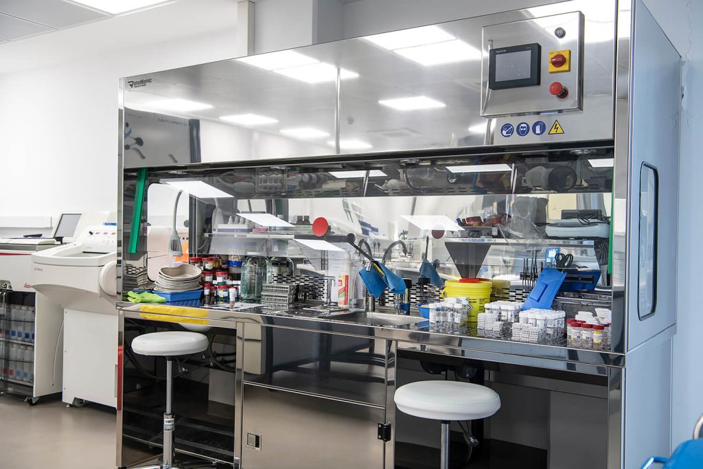
Grossing Workstation
This is a workstation that has been specifically designed for the macroscopic processing of tissues with a large working area that is resistant to chemicals, with a rinsing system for the collection of fluids and with filters that keep the formaldehyde fumes away from the user, creating a safer and healthier work place.
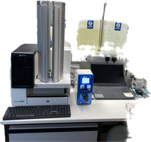
Cassette and Slide Laser Printer
In our department, we have an innovative and fully autonomous system which is able to laser print codes onto glass slides and cassettes. Printing is controlled by internal machine software. This equipment offers permanent and processing resistant marking. It ensures our specimens are accurately labelled and eliminates errors in tissue entanglement.
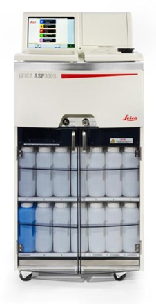
Automatic Fully Enclosed Tissue Processing Machine Leica ASP300S
We feel comfortable knowing that we have quality tissue processing that meets our department's high standards.
Our fully enclosed automatic tissue processor performs tissue dehydration, clearing, and paraffin impregnation. It combines proven technology and modern design, provides high reliability, and allows many tissue samples to be safely processed in each run.
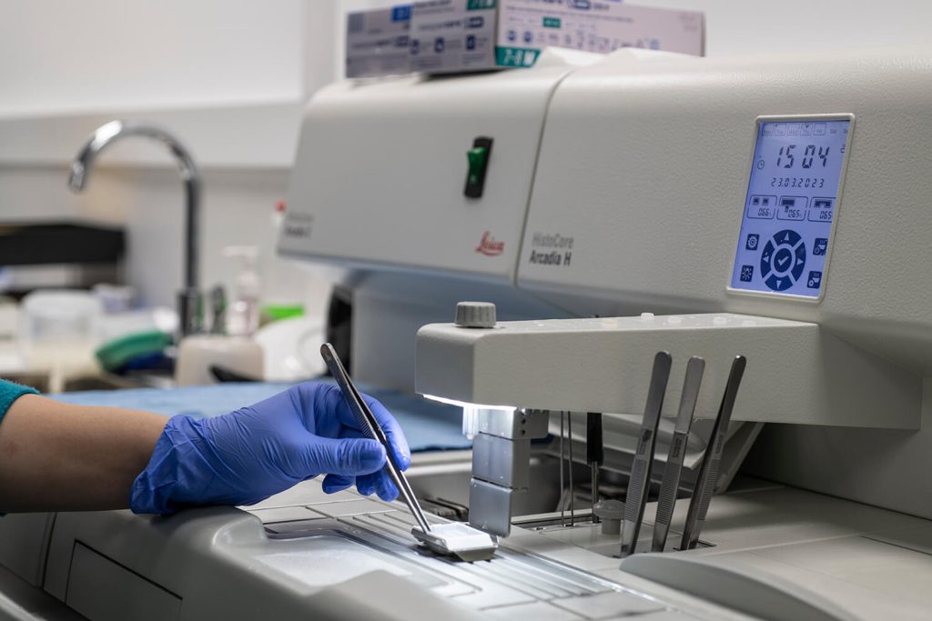
Paraffin Embedding Tissue Station
To further improve tissue preservation and allow samples to be stored safely for years, for future diagnostic or research purposes, tissues which have been chemically fixed with formaldehyde can be embedded in paraffin. The paraffin embedded tissue station enables us to produce paraffin embedded tissue blocks. The station incorporates two separate components: a heated paraffin dispensing system and a cold plate by which we ensure the correct tissue orientation and create the ideal block shape. The paraffin embedded unit features a digital programmable interface which enables us to program individual temperature settings for the paraffin reservoir and the work surface.
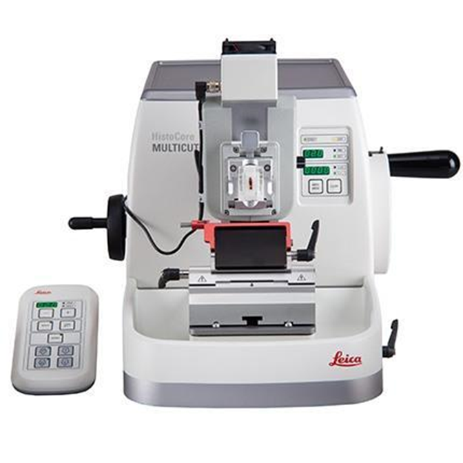
Microtomes
An accurate diagnosis relies largely on precise tissue sections. We use modern semiautomatic rotary microtomes to cut thin tissue slices for observation under the microscope. The cut tissue is floated over a water bath to eliminate wrinkles and distortion, then transferred to a glass slide. Next, the slides are heated in incubators and are ready for staining.
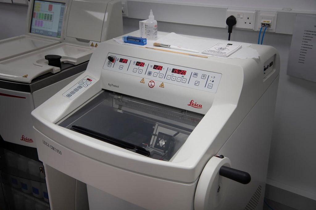
Cryostat
When a Clinician (especially a surgeon) requests an intraoperative consultation, the Department of Pathology must be alert and ready. Therefore, a pathologist is often required to make rapid diagnostic decisions and to help determine, for example, how much tissue should be removed when excising cancerous tissues.
With the cryostat, which uses cryogenic temperatures, we can cut tissue sections, stain, and examine them under a microscope in a very short period (within approximately 15 minutes), providing a very quick diagnosis for a variety of diseases and medical conditions. With this procedure, we help our colleagues decide on further surgical interventions.
In our department, we use the most modern cryostat, which can meet requests for immediate results by quickly, accurately, and safely cutting and preserving frozen sections of tissue. Moreover, the speed and versatility of our cryostat help us collect and analyze more specimens each day.
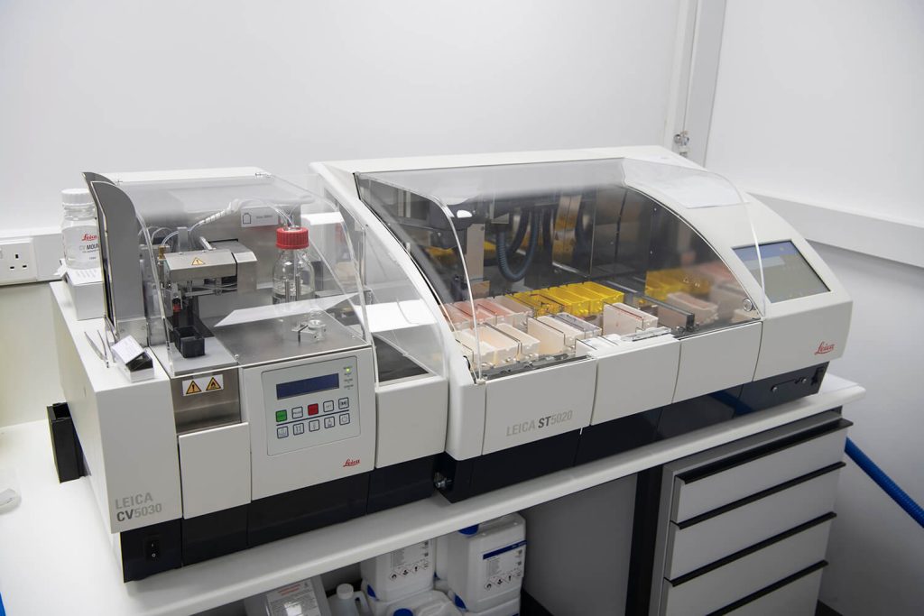
Automatic Slide Staining Machine and Automated Glass Coverslip
This is a workstation automatic slide stainer, which is used for diagnostic and research purposes, and provides a broad view of various cellular and tissue characteristics in formalin-fixed, paraffin-embedded, or freshly frozen tissue. The Haematoxylin-Eosin stain is an essential routine process through which the cell nucleus and cytoplasm are stained with different colours. The information gathered through this staining process allows our pathologists to observe changes within cells and tissues in great detail, enabling them to make a diagnosis.
The automatic slide staining machine used in our department performs different staining protocols (e.g., protocols for special stains) simultaneously, offers high levels of stain quality, reliability, high throughput due to the innovative design and high capacity of lot of cuvette stations as well as workflow efficiency.
Our automated glass coverslipper produces slides with superior optical quality for reliable long-term storage. It can handle a large variety of slide racks and provides consistent, reliable, and fast coverslipping enabling us to not delay the diagnostic procedure.
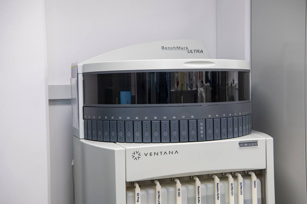
Autostainer for Immunohistochemistry (IHC) and Chromogenic In Situ Hybridization (CISH)
Pathologists often rely on immunohistochemistry (IC) and in situ hybridization (ISH) slides for a definitive diagnosis.
Immunohistochemistry (IHC) has been an invaluable tool for the detection, localization, and quantification of antigens in preserved tissue for diagnostic and research purposes. IC is crucial for the accurate classification of tumors, the determination of a metastatic tumor's site of origin and for the detection of isolated cells or foci of tumor cells which are inconspicuous on routine Haematoxylin-Eosin and Eosin staining. It is increasingly being used by pathologists to provide predictive and prognostic information for patients with a variety of cancer types, that may aid in determining the most appropriate treatment for that individual.
Chromogenic in situ hybridization (CISH) is a cytogenetic technique that is frequently applied to cancer tissues to assess gene amplification, chromosomal rearrangements, and fusions. Apart from cancers, CISH has also been useful in detecting virus infections.
Our department uses an instrument with the ability to work both IC and CISH methods simultaneously, thus allowing both more kinds of tests to be performed, and more tissues to be processed.
Using this system increases the flexibility, quality, and productivity of our department with the ultimate impact of providing reliable and accurate diagnoses.

Microscopes
Pathologists use microscopes for diagnostic or research purposes. The microscopic examination of tissues often requires long working hours.
With the help of a microscope, pathologists can easily view the magnified images of all types of cells and tissues, extract very small details, and see abnormalities that they would not be able to see with their naked eye. Moreover, they can interpret the results of special stains (histochemical, immunohistochemical, ISH) for an integrated diagnosis, to provide information for prognosis or treatment predictions, and for research purposes.
Therefore, a precise microscopic examination is of paramount importance for the diagnosis and treatment of a patient.
Microscopes used in our department are of innovate technology and design, offer excellent ergonomics for comfortable operation and high-quality optics for high colour reproducibility. Modular options enable different observation styles like brightfield or darkfield.
Moreover, they are equipped with a camera so subtle colour variations can be captured in images for reporting and archiving.