Heart Valve Disease
Overview
In Europe, it is estimated that 13% of people aged 75 and older have valvular heart disease (VHD). Prevalence increases markedly after age 65. Severe VHD leads to the deterioration of the heart’s function. This may require hospitalization and intervention, and can be terminal.
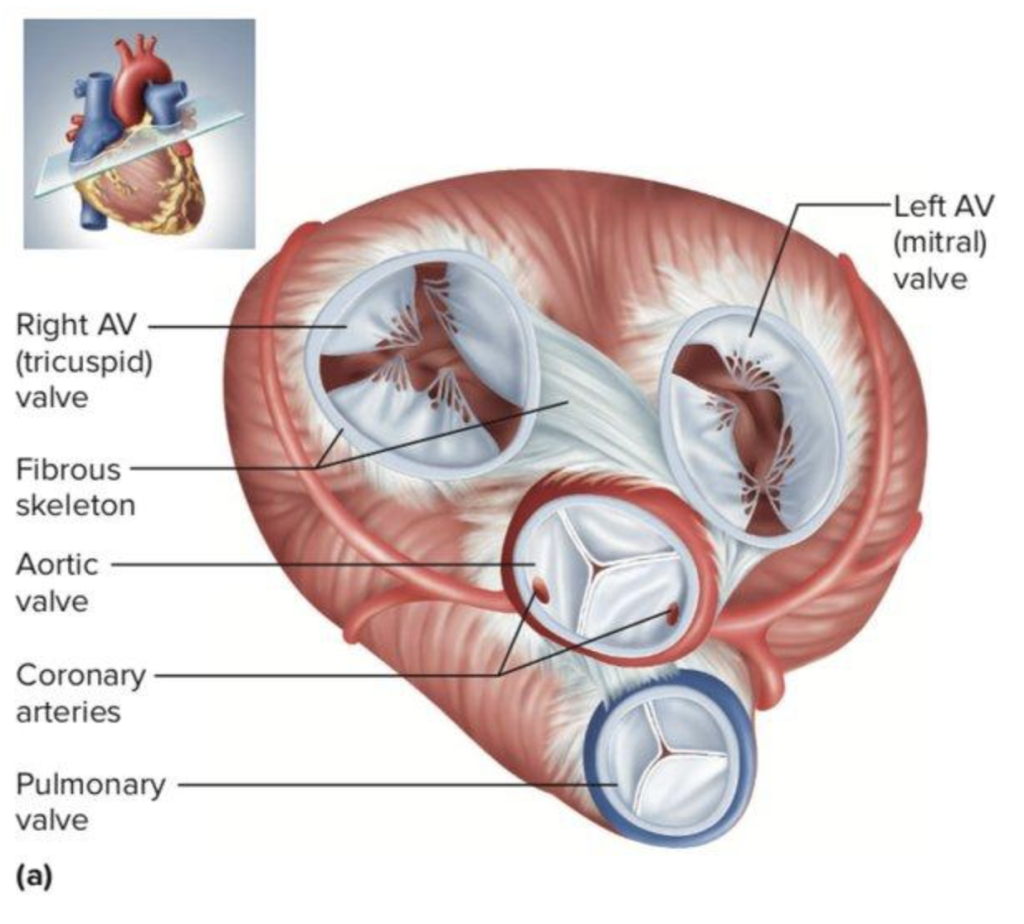
There are 4 valves in the heart: aortic, mitral, tricuspid, and pulmonary. One or more of these can be affected by the disease. In western countries, degenerative aortic valve disease is most common, followed by mitral valve disease. Tricuspid and pulmonary valve disease are less prevalent. When it comes to calcific aortic valve disease, Cyprus presents the largest age standardized death rate in Europe with 8.20 per 100,000 persons, and it is among the countries where this trend is increasing.
The main risk factors for cardiac valve disease are age, hypertension (high blood pressure), hypercholesterolemia (high cholesterol), diabetes, infections, smoking, a history of heart attack, and congenital heart conditions.
Symptoms
The most common symptoms of heart valve disease include shortness of breath, fatigue, dizziness, fainting, chest pain, swelling of the ankles, and an irregular heartbeat (palpitation).
These symptoms can occur at any time, such as during exertion or when resting. If you experience any of these symptoms, it is important that you contact your medical team immediately.
Diagnosis
If you are experiencing any of the above symptoms, after a consultation, your medical team will recommend further testing. This can include:
An electrocardiogram (ECG), which measures the electrical activity of your heart. It can check whether your heart is beating regularly and show the pace of your heartbeat. It can also show whether you are having or have had a heart attack.
An echocardiogram, which uses ultrasound to create images of the beating heart. An echocardiogram can assess if the heart chambers and valves are healthy or damaged.
A cardiac magnetic resonance imaging (CMR), which uses powerful magnetic fields to build a detailed image of the cardiovascular system. This is a non-invasive test which can provide lots of information on the condition of your heart. It can help your medical team evaluate the anatomy and function of the heart chambers, heart valves, size and blood flow through major vessels, and the surrounding structures, such as the pericardium. It is used to diagnose a variety of cardiovascular disorders, including anomalies like valve disease, tumors, infections, and inflammatory conditions.
Since valvular disease of the heart may also be associated with coronary heart disease, your medical team may also recommend some further testing, including a Cardiac CT scan and Cardiac Catheterization.
A Cardiac CT scan can visualize the details of the heart, show any calcium deposits which narrow arteries, and any blockages in the heart arteries. Contrast, which is administered intravenously, helps create detailed pictures of the arteries. If contrast is administered, the test is called a CT coronary angiogram.
A cardiac catheterization and angiogram test can show the blockages in your arteries. During a cardiac catheterization, an interventional cardiologist gently inserts a very thin, flexible tube (catheter) into a blood vessel, which is accessed through the wrist or groin. The catheter is then guided to the heart, with the help of X-ray images. Dye, which flows through the catheter, helps your blood vessels show up more clearly on the X-ray images and outlines any blockages.
Treatment
Valvular heart disease is mostly treated by repairing or replacing the damaged valve. After a consultation, your cardiac surgeon will arrange all the necessary tests prior to the operation.

Valve repair is a surgical procedure that allows the patient to keep his native valve. It can only be performed for certain cases of valve disease. If it’s not possible to repair the native valve, then your cardiac surgeon will replace it with a new prosthetic one. Sometimes a minimally invasive heart surgery, which involves smaller incisions than those made for open-heart surgery is doable.
During the operation, the patient undergoes general anesthesia. The cardiac surgeon will then repair the affected valve by modifying it to make it functional again (valvuloplasty). After the operation, the patient will stay in our cardiac intensive care ward for 1-2 days while they recover, then they will be transferred to our regular ward. The average hospital stay after a valve repair is 5-7 days, but this varies depending on the patient’s pre-operative clinical condition.
Valve replacement surgery is recommended when repair is not possible. Sometimes a minimally invasive heart surgery, which involves smaller incisions than those made for open-heart surgery is doable. Also, sometimes a percutaneous transcatheter valve replacement is indicated and is performed from a catheter through the blood vessels of the groin. This is the least invasive form of valve replacement surgery and is suitable for only a specific group of patients.
There are 2 types of prosthetic valves that can be used (biological or mechanical) during valve replacement surgery. Both have different features, and your cardiac surgeon will help you to choose which will be the best for you. During the operation, the patient undergoes general anesthesia. The cardiac surgeon will then replace the affected valve with a new prosthetic one. After the operation, the patient will stay in our cardiac intensive care ward for 1-2 days while they recover, then they will be transferred to our regular ward. The average hospital stay after a valve replacement is 5-7 days, but this varies depending on the patient’s pre-operative clinical condition.
Why GMI
At the GMI, we have a state-of-the-art facility for patients affected by cardiac diseases, which includes our Cardiovascular Diagnostic Center, Cardiology Catheterization Laboratory, Cardiac Surgery Theatre, Hybrid Theatre and Cardiac Intensive Care Unit.
Our team of internationally recognized heart doctors (cardioradiologists, cardiologists, cardiac surgeons and cardioanaesthetist) will guide you through the whole process, from your diagnostic work-up to your treatment and post-treatment care. We are committed to providing the best treatment options to each of our patients. The GMI team will never offer a simple “one size fits all” approach to any patient. We believe each patient’s case is as individual as they are and strive to find the best solution for each of our patients, taking their specific case and diagnosis, their lifestyle, and choices into account.


