Thoracic aorta diseases
Overview
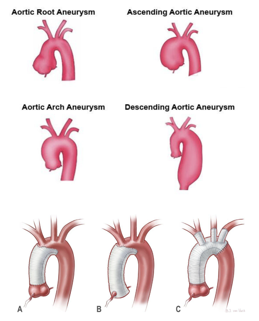
The aorta is the largest artery in the human body and it carries blood from the heart to the rest of the organs. When the aorta significantly widens, it’s called aortic aneurysm. Globally, around 5.3 people per 100,000 have thoracic aortic aneurysms (TAA) each year. Depending on where the thoracic aneurysm occurs, it is either an aortic root or ascending aortic aneurysm, which is the most common type (≈60%), followed by an aneurysm of the descending aorta (≈35%) and of the aortic arch (<10%). Thoracic aortic aneurysms occur more often in men, and can have life threatening complications including a rupture of the aorta or a tear between the layers of the aorta’s wall, which is called an aortic dissection.
The main risk factors for thoracic aortic disease include a family history of the disease, age, (where growing older increases your risks), high blood pressure (hypertension), chronic obstructive pulmonary disease (COPD), smoking, inflammatory diseases, having a bicuspid aortic valve, and genetic conditions such as Marfan syndrome and Ehlers-Danlos syndrome.
Symptoms
Aortic aneurysms are a silent disease, often growing slowly and without symptoms. They are usually discovered accidentally, however when there is a dissection or an impending rupture of the aorta, you may feel a sharp pain in the upper back that spreads downwards, chest pain, and you may lose consciousness. If the aortic aneurysm extends to the root of the aorta, this can impair the aortic valve, leading to a valve regurgitation, and causing shortness of breath.
Diagnosis
Thoracic aortic aneurysms are found through imaging tests. If you have any symptoms of a TAA, or are considered high risk, your medical team may recommend you undergo imaging studies. TAA’s are most often discovered when imaging studies are conducted for other reasons. Cardiac magnetic resonance imaging (CMR), thoracic computed tomography (CT) and echocardiograms are the best ways to diagnose a TAA.
Cardiac Magnetic resonance imaging (CMR) uses powerful magnetic fields to build a detailed image of the cardiovascular system. This is a non-invasive test which can provide lots of information on the condition of your heart. It can help your medical team evaluate the anatomy and function of the heart chambers, heart valves, size and blood flow through major vessels, and the surrounding structures such as the pericardium. It is used to diagnose a variety of cardiovascular disorders.
An aortic CT scan can visualize the details of the aorta. Contrast, which is administered intravenously, helps create detailed pictures of the arteries.
An echocardiogram uses ultrasound to create images of the beating heart. An echocardiogram can assess if the heart chambers and valves are healthy or damaged.
Treatment
Thoracic aortic aneurysms are treated by surgically replacing the affected part of the aorta.

After a consultation, a cardiac surgeon will arrange all the necessary tests prior to the operation. During the operation, the patient undergoes general anesthesia. The surgeon will repair the affected area of the aorta by replacing it with a synthetic tubular prosthesis. After the operation, the patient will stay in our cardiac intensive care ward for a few days while they recover, then they will be transferred to our regular wards. The average hospital stay after aortic surgery is 7-10 days, but this varies depending on the patient’s pre-operative clinical condition.
Why GMI
At the GMI, we have a state-of-the-art facility for patients affected by cardiac diseases, which includes our Cardiovascular Diagnostic Center, Cardiology Catheterization Laboratory, Cardiac Surgery Theatre, Hybrid Theatre and Cardiac Intensive Care Unit.
Our team of internationally recognized heart doctors (cardioradiologists, cardiologists, cardiac surgeons and cardioanaesthetist) will guide you through the whole process, from your diagnostic work-up to your treatment and post-treatment care. We are committed to providing the best treatment options to each of our patients. The GMI team will never offer a simple “one size fits all” approach to any patient. We believe each patient’s case is as individual as they are and strive to find the best solution for each of our patients, taking their specific case and diagnosis, their lifestyle, and choices into account.


