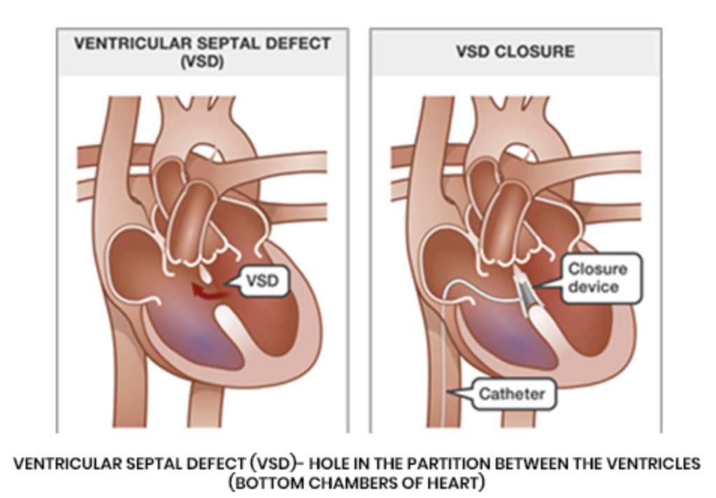
Ventricular Septal Defect
Overview
Lorem ipsum dolor sit amet, consectetur adipiscing elit. Ut elit tellus, luctus nec ullamcorper mattis, pulvinar dapibus leo. Lorem ipsum dolor sit amet, consectetur adipiscing elit. Ut elit tellus, luctus nec ullamcorper mattis, pulvinar dapibus leo. Lorem ipsum dolor sit amet, consectetur adipiscing elit. Ut elit tellus, luctus nec ullamcorper mattis, pulvinar dapibus leo. Lorem ipsum dolor sit amet, consectetur adipiscing elit. Ut elit tellus, luctus nec ullamcorper mattis, pulvinar dapibus leo. Lorem ipsum dolor sit amet, consectetur adipiscing elit. Ut elit tellus, luctus nec ullamcorper mattis, pulvinar dapibus leo. Lorem ipsum dolor sit amet, consectetur adipiscing elit. Ut elit tellus, luctus nec ullamcorper mattis, pulvinar dapibus leo. Lorem ipsum dolor sit amet, consectetur adipiscing elit. Ut elit tellus, luctus nec ullamcorper mattis, pulvinar dapibus leo. Lorem ipsum dolor sit amet, consectetur adipiscing elit. Ut elit tellus, luctus nec ullamcorper mattis, pulvinar dapibus leo. Lorem ipsum dolor sit amet, consectetur adipiscing elit. Ut elit tellus, luctus nec ullamcorper mattis, pulvinar dapibus leo. Lorem ipsum dolor sit amet, consectetur adipiscing elit. Ut elit tellus, luctus nec ullamcorper mattis, pulvinar dapibus leo. Lorem ipsum dolor sit amet, consectetur adipiscing elit. Ut elit tellus, luctus nec ullamcorper mattis, pulvinar dapibus leo. Lorem ipsum dolor sit amet, consectetur adipiscing elit. Ut elit tellus, luctus nec ullamcorper mattis, pulvinar dapibus leo.
Symptoms
Signs and symptoms vary depending on the size of the hole and if you have any other associated heart defects.
Symptoms in a baby or child may include poor eating, failure to thrive, fast breathing or breathlessness and tiring easily.
Patients with small VSDs can be asymptomatic. However, depending on the size and type of defect, you may experience shortness of breath, fatigue, or an irregular or slow heartbeat. VSDs can also lead to complications such as aortic regurgitation (leaking of one of the heart valves), heart failure, pulmonary hypertension, and endocarditis (infection of the heart).
Diagnosis
VSDs often cause a heart murmur that your doctor can hear using a stethoscope. If you or your child have a murmur or any symptoms of a VSD, your medical team may recommend imaging studies. These include an echocardiogram, a transesophageal echocardiogram, and a cardiacmagnetic resonance imaging (CMR).
An echocardiogram uses ultrasound to create images of the beating heart. An echocardiogram can assess if the heart chambers and valves are healthy or damaged.
Transesophageal echocardiograms give a more detailed picture of the heart structures. Your doctor will use a small probe, which is inserted through the mouth down into the tube that connects your mouth to your stomach (the esophagus). Patients undergoing this exam will be given a light sedation to help make them as comfortable as possible.
A cardiac magnetic resonance imaging (CMR) uses powerful magnetic fields to build a detailed image of the cardiovascular system. This is a non-invasive test which can provide lots of information on the condition of your heart. It can help your medical team evaluate the anatomy and function of the heart chambers, heart valves, size and blood flow through major vessels, and the surrounding structures such as the pericardium. It is used to diagnose a variety of cardiovascular disorders, including anomalies like VSD.
Treatment
In some cases, when the hole is small in size, only monitoring is required. If you experience shortness of breath, heart failure, or pulmonary hypertension, the cardiologists will recommend treatment.
It is possible to close the hole through a percutaneous procedure, where an interventional cardiologist inserts a small wire in the groin and guides a device between the heart chambers. The device is then opened up, which closes the hole.
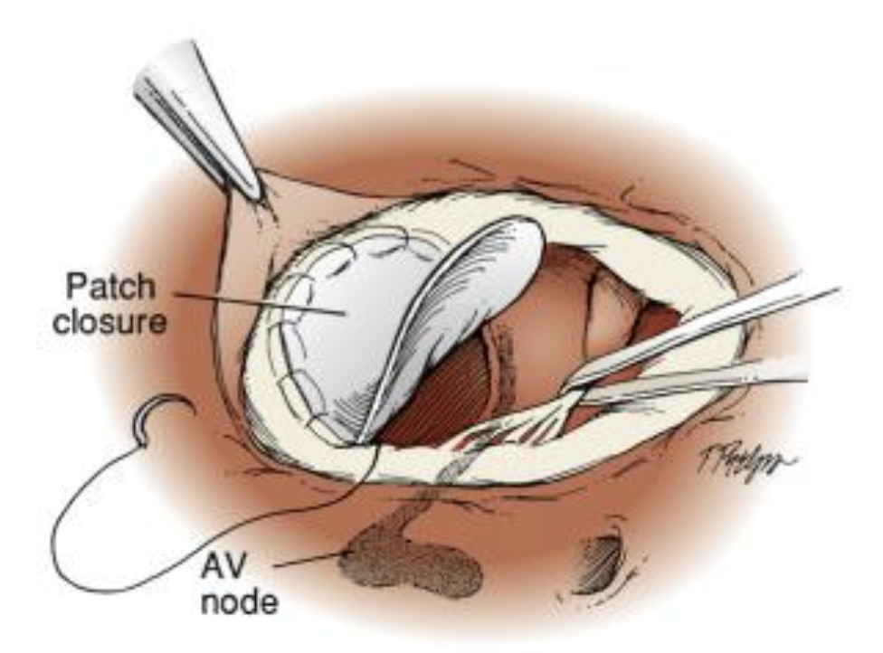
In more complex cases, when it is not possible to use a closure device, surgical repair is recommended. After a consultation, a cardiac surgeon will arrange all the necessary tests prior to the operation. During the operation, the patient undergoes general anesthesia. The surgeon then patches the hole using a biological membrane, which is sutured between the heart chambers. After the operation, the patient will stay in our cardiac intensive care ward for a few days while they recover, then they will be transferred to our regular ward. The average hospital stay after a surgical VSD closure is 7-10 days, but this varies depending on the patient’s pre-operative clinical condition.
Why GMI
At the GMI, we have a state-of-the-art facility for patients affected by cardiac diseases, which includes our Cardiovascular Diagnostic Center, Cardiology Catheterization Laboratory, Cardiac Surgery Theatre, Hybrid Theatre and Cardiac Intensive Care Unit.
Our team of internationally recognized heart doctors (cardioradiologists, cardiologists, cardiac surgeons and cardioanaesthetist) will guide you through the whole process, from your diagnostic work-up to your treatment and post-treatment care. We are committed to providing the best treatment options to each of our patients. The GMI team will never offer a simple “one size fits all” approach to any patient. We believe each patient’s case is as individual as they are and strive to find the best solution for each of our patients, taking their specific case and diagnosis, their lifestyle, and choices into account.