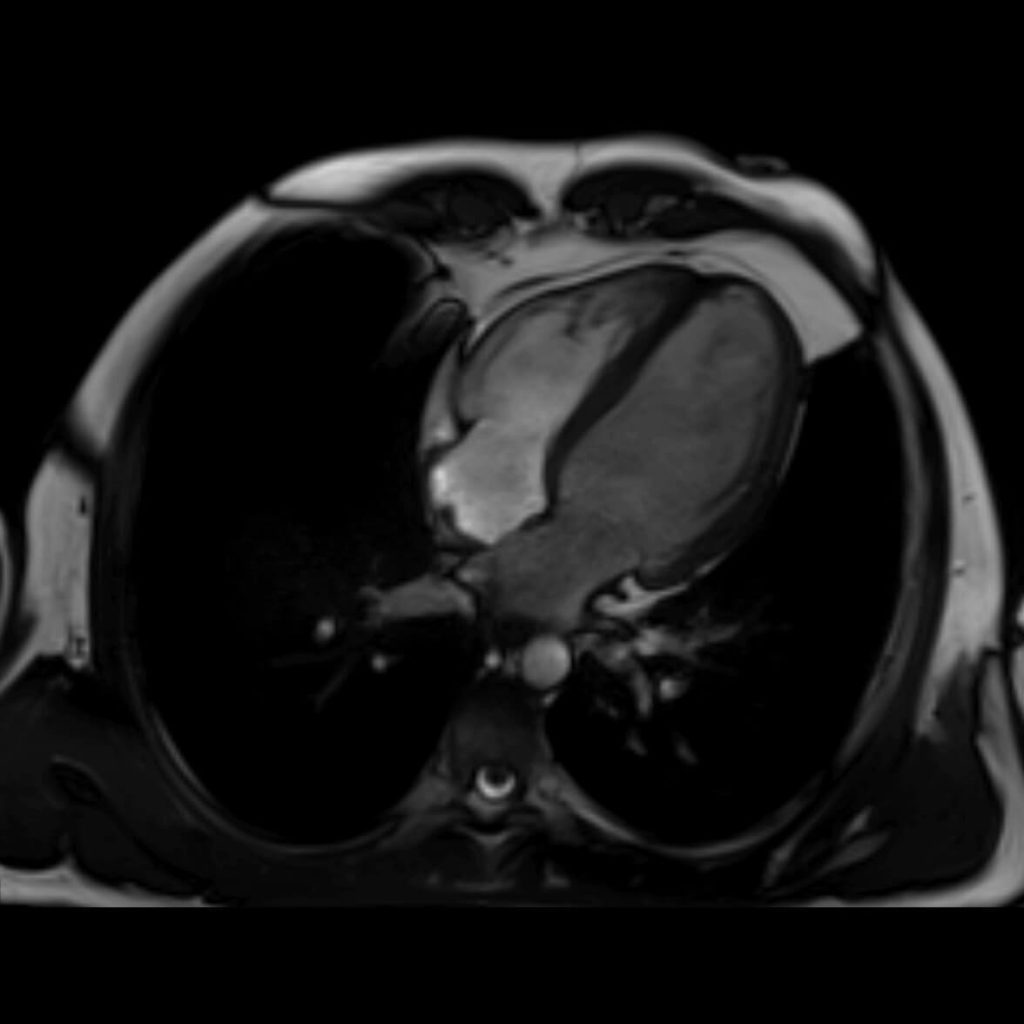
Basic Examinations
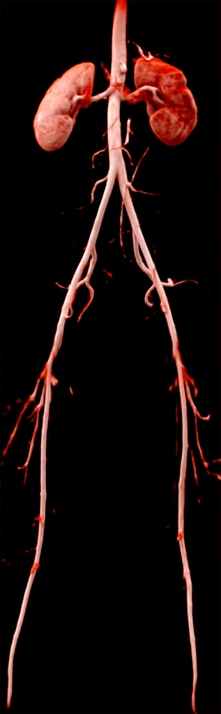
Angiography / Venography - (With or without the use of intravenous contrast agent)

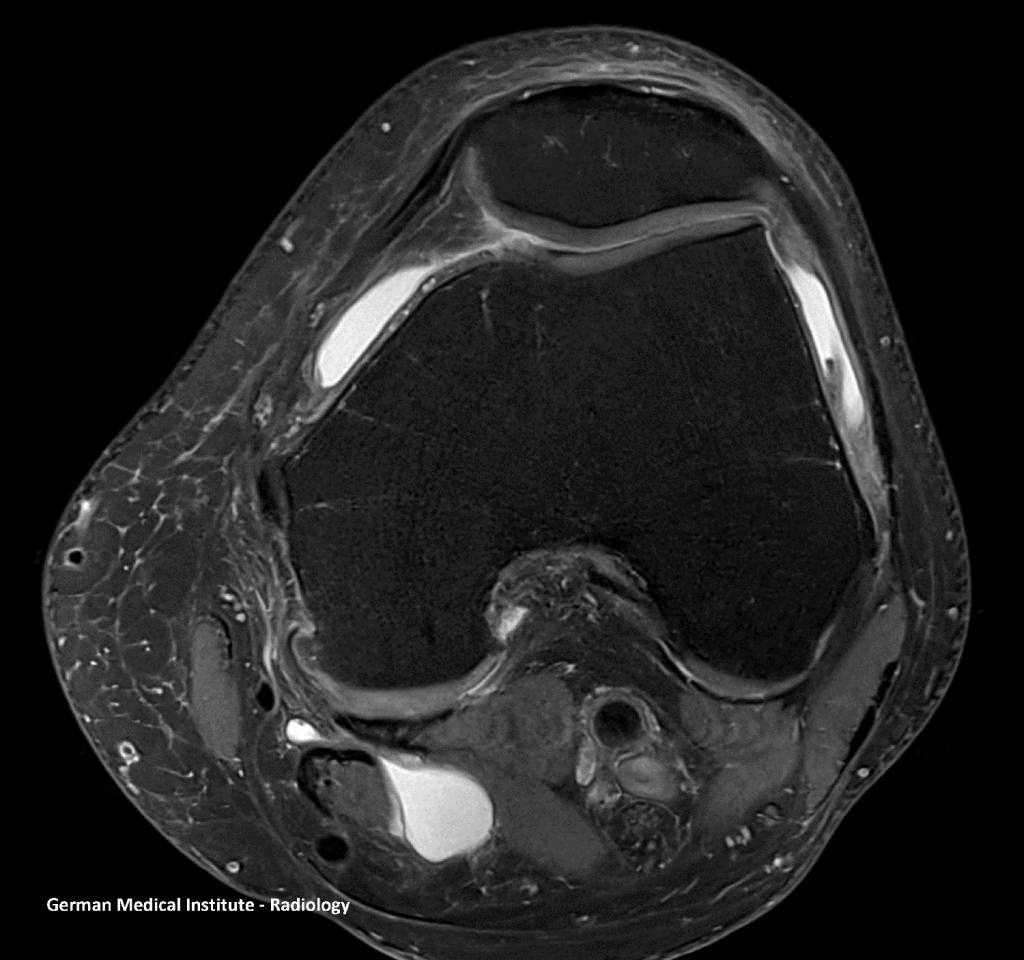
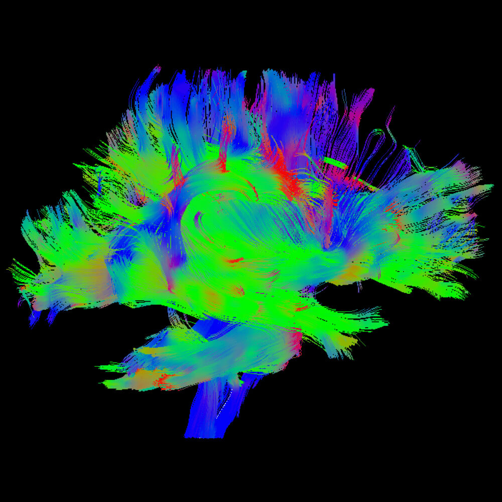
Specialised Examinations
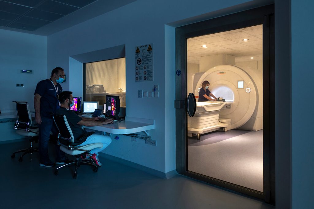
Preparing examinees for MRI scans
Basic Examinations
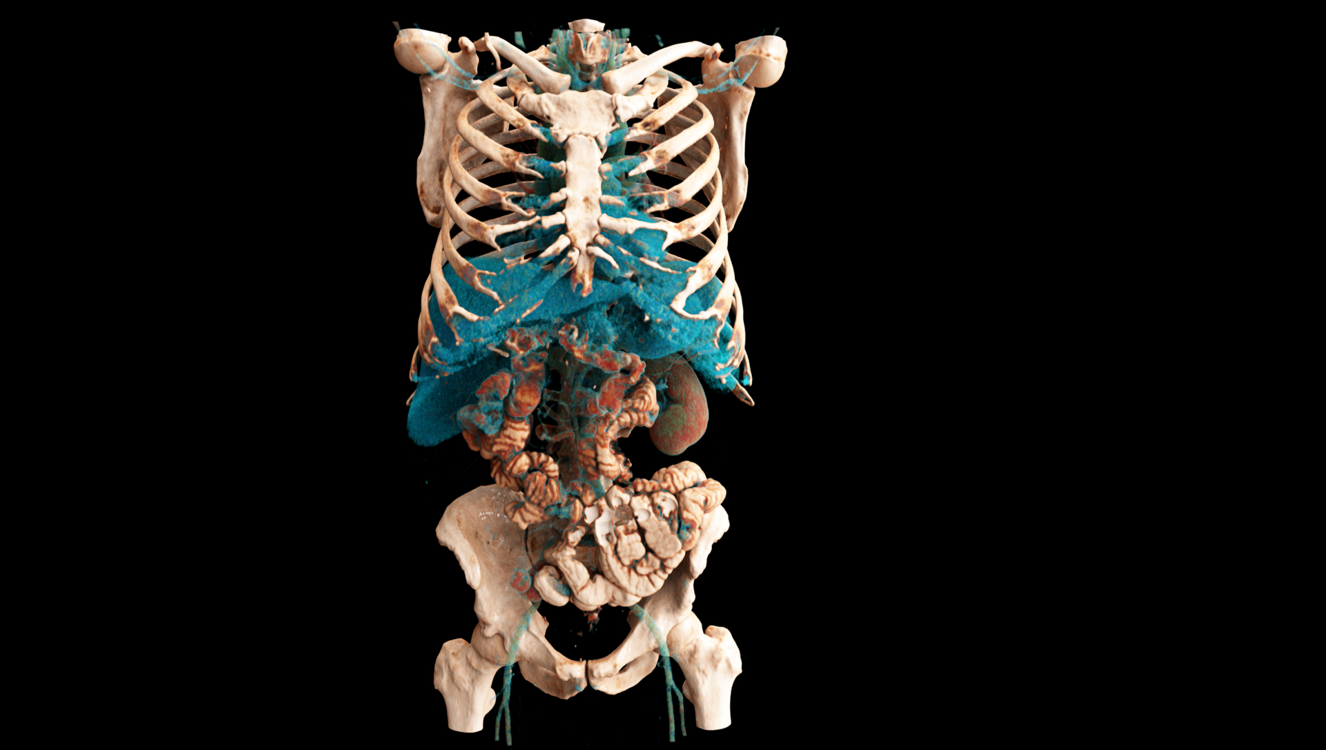
Angiography
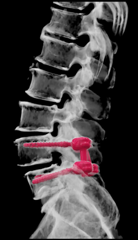
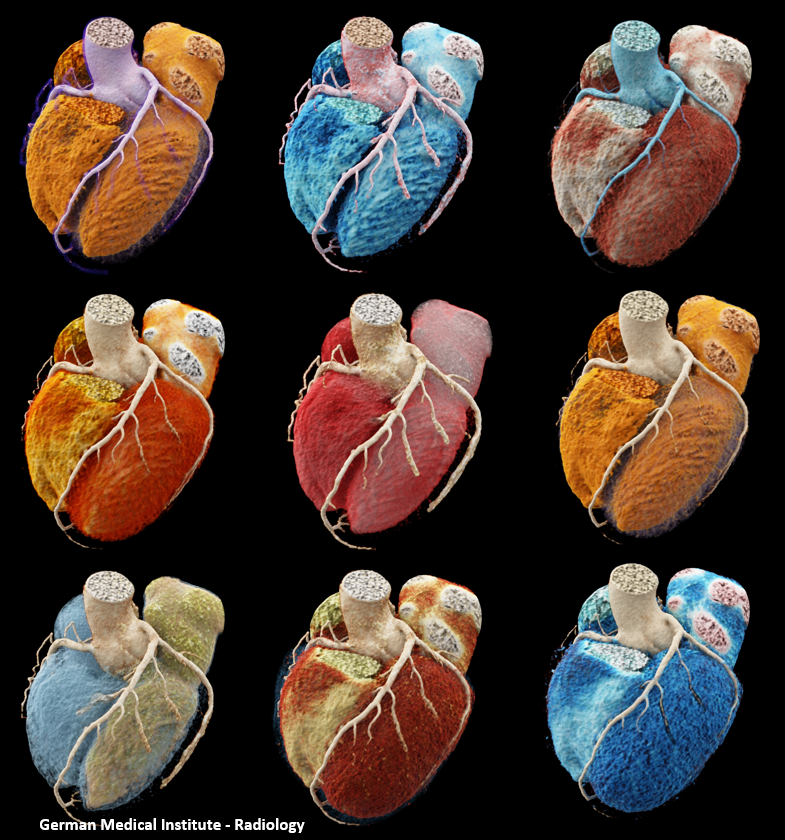
Specialised Examinations
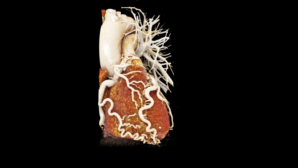
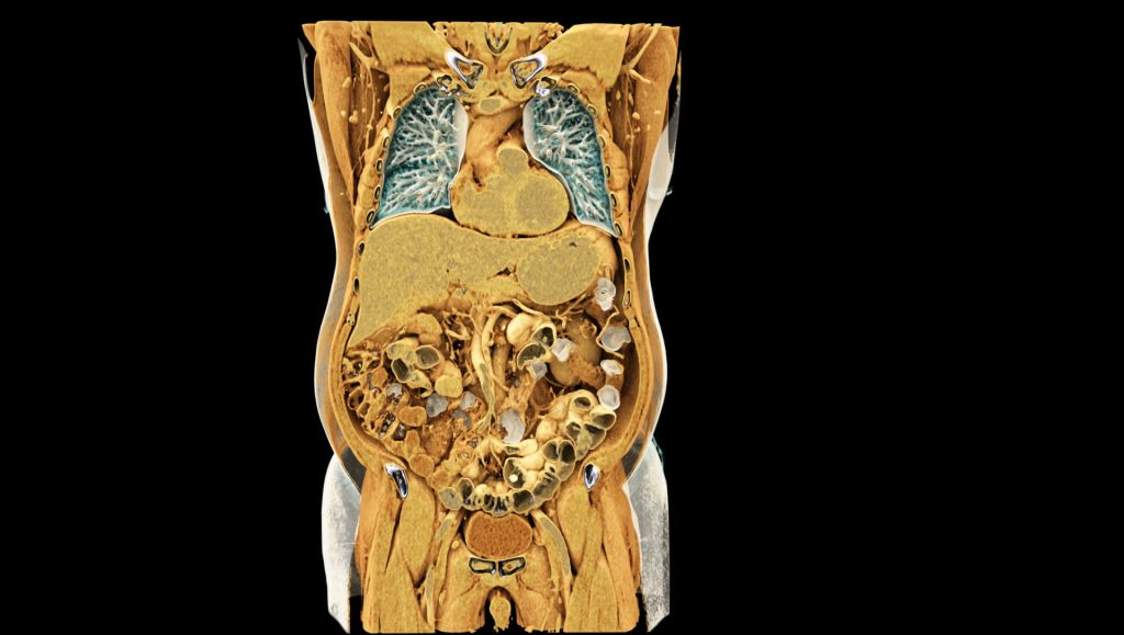
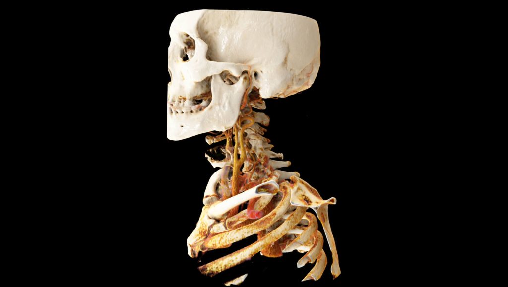
Preparing examinees for CT scans
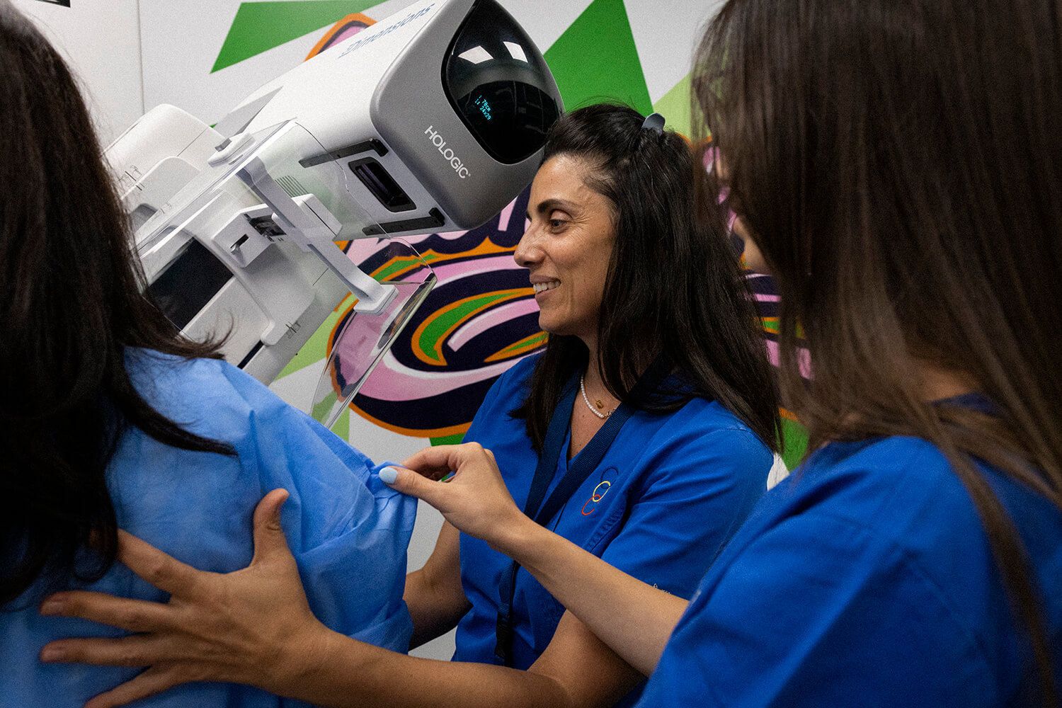
Basic exams for screening and/or diagnosis