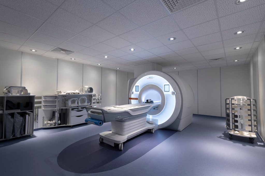
Magnetic Resonance Imaging
The two MRI units, operating at 1.5 and 3 Tesla, offer a wide range of conventional and specialised examinations. Both systems make use of advanced artificial intelligence and deep learning image reconstruction systems, in conjunction with innovative imaging methodologies. The synergy of these factors expedites the examination processes while maintaining impeccable image quality. The patient-centric approach offers the examinee convenience in accessibility and comfort during the examination, accommodating both claustrophobic or larger individuals, with a maximum allowable examinee weight reaching 250kg.
Siemens Magnetom VIDA 3T with AI technology (Deep Resolve)
The centre is equipped with the most advanced Siemens MRI scanner, Magnetom VIDA 3T. This system represents the first MRI scanner with biometric technology, enabling wireless tracking of patient biometrics without the presence of cables. In conjunction with its large 70 cm bore diameter and spacious room, it cultivates a more relaxed and pleasant environment for the patient.
The system is equipped with multi-channel coil technology capable of capturing images using up to 64 channels, which represent the highest channel count presently available in Cyprus. This facilitates faster examinations and ensures exceptional image quality. The equipment is complemented by an array of coils featuring a large number of independent channels, such as the Biomatrix Head/Neck 64 brain coil and the Biomatrix 72 spine coil, both of which stand at the forefront of coil technology from Siemens. The vast selection of coils configuring the MR system permits the execution of a wide range of examinations with flexible combinations, all without subjecting examinees to discomfort due to the need for repositioning.
The equipment is complemented by Siemens’ state-of-the-art software composition, featuring image reconstruction capabilities with the artificial intelligence technology, deep learning Deep Resolve. This represents the cutting-edge of MR imaging, offering high-resolution imaging and reduced examination duration. The software is supported by sophisticated post-processing and analysis software designed for neurological, oncological, cardiac, angiography, and various other advanced examinations.
GE Signa HDxt +SW 1.5T with AI technology (AirRecon DL)
The General Electric MRI system is equipped with a comprehensive set of coils and accessories, enabling a broad spectrum of diagnostic examinations, MRI-guided interventional procedures, and diagnostic exams under anaesthesia. The MRI suite offers ample space, enabling patients, in specific examinations, to recline with their head positioned outside the bore, and provides the option to enjoy their preferred choice of music during the procedure.
Upgraded to the latest software version, it provides image reconstruction capabilities with artificial intelligence through Air Recon DL. This technology opens new horizons in MR imaging, providing enhanced image quality at accelerated speeds. The software offers image post-processing features for advanced exams, and enables reconstructions that enhance the diagnostic value of the examination.
Computed Tomography
The Department of Diagnostic and Interventional Radiology is equipped with two CT scanners. One of these is a single-energy system featuring 16 detector rows, while the other is a state-of-the-art, next-generation system CT scanner with dual-source, dual-energy capabilities and 384 detector rows.
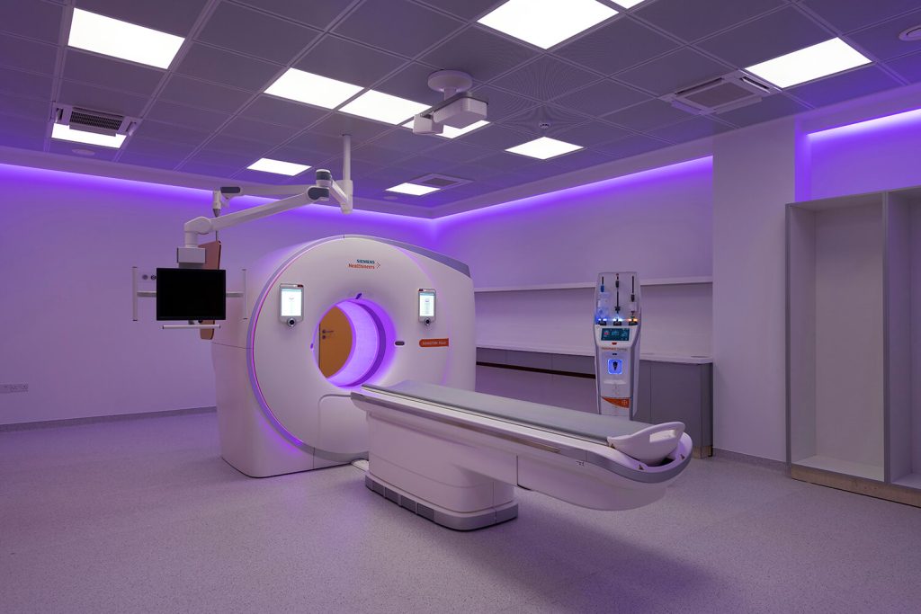

Siemens Somatom Force - Dual Source - Dual Energy
The department houses the state-of-the-art third generation 384 (2×192) slice CT scanner, which is unique in the Cyprus-Greece region. This system encompasses two X-ray tubes mounted within the gantry at an angle of approximately 90 degrees. Notably one of the tubes features an additional tin filter, designed to selectively eliminate lower-energy photons, thereby achieving a substantial reduction in absorbed dose. With its advanced technology the scanner offers superior temporal resolution when contrasted with its single-tube counterparts, with a temporal resolution of 66 ms and a scanning speed of up to 737 mm/s. This translates into expanded capabilities, particularly in cardiac imaging, where the whole heart can be captured within 0.3 seconds. The dual-source CT scanner excels in generating high-quality images over a wide range of heart rates. Moreover, the system incorporates a 3D camera, ensuring precision in patient alignment at the isocenter of the scan. Another advantage of the X-ray tubes of this system is the enhanced power they provide, enabling improved imaging for larger patients. This feature also allows for rapid imaging for large coverages, which is crucial in emergency situations.
Furthermore, the simultaneous utilisation of dual X-ray beams operated at different energies allows the differentiation of tissues based on their atomic numbers and thus their unique absorption profiles, a feature known as material decomposition. This capability is relatively new in the field and continually unveils novel clinical applications. Notable examples include the replacement of initial non-contrast scans with virtual unenhanced scans, the identification of urinary stone composition, bone elimination in angiography procedures, and the identification of uric acid and bone marrow oedema. The software suite is complemented by dedicated post-processing and analysis tools tailored for neurological, oncological, cardiac, angiography, and other specialised examinations.
CT Optima 580/Optima 580 RT
The General Electric 16-slice CT scanner features a large 80 cm bore diameter for greater patient comfort. An additional attribute of this system is its capacity to incline the entire gantry, rendering it ideal for a wide range of interventional procedures. Beyond the broad spectrum of interventional procedures, the system also serves the Department of Radiation Oncology for patients’ radiotherapy plans. Using the three-dimensional images generated by the CT scanner, allows for precise contour of the target planning volumes, a prerequisite for high quality treatment planning.
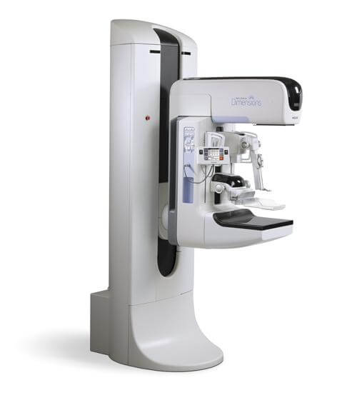
Digital Mammograph
The diagnostic department is equipped with the state-of-the-art digital mammography system from Hologic, Selenia 3Dimensions. The American company has left its mark on breast imaging, as it is the first to develop technology for three-dimensional imaging through digital tomosynthesis. This technique has the capability to characterise even small benign or malignant findings with greater sensitivity. In combination with the high-resolution detector it features, it improves the detection capabilities of architectural disorders, subtle masses even in dense breasts.
The system enables more precise imaging while minimising radiation exposure by means of automatic radiation dose adjustment tailored to the size of the breast and its composition. Furthermore, It enhances comfort for the examinees by incorporating a specially designed compression paddle, ensuring a more comfortable breast positioning, along with implementing techniques aimed at pain reduction during compression.

The equipment is accompanied by a dedicated workstation for mammography examinations, with the upgraded SecureView software by Hologic, which provides workflow customisation and automation, allowing image post-processing for tomosynthesis and 2D synthetic image reconstruction without additional radiation exposure.
The diagnostic tools offered by the breast department are enhanced by Hologic’s stereotactic biopsy system. Leveraging advanced automated robotic technology, this system stands as one of the modern biopsy methods. Employing 3D technology, it enables the precise and expeditious targeting of lesions with a needle. Utilising a vacuum-assisted biopsy mechanism, it efficiently acquires large tissue samples, thereby ensuring precise lesion targeting. The same stereotactic approach is applied for accurate preoperative placement of localisation hookwire.
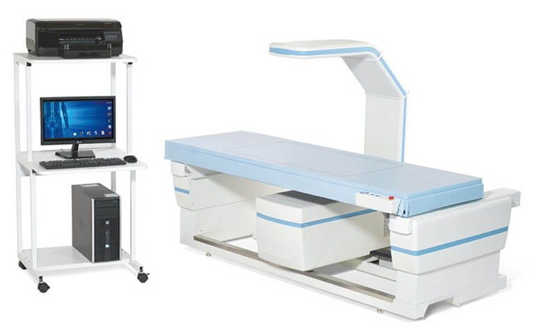
DEXA
The diagnostic department has the modern bone mineral density system from Hologic, DEXA Horizon Wi. This system is capable of emitting photon beams at two energies, through advanced fan-beam technology, rendering it exceptionally fast, stable and of low radiation dose. The equipment features a high-resolution ceramic detector, accompanied by a high-performance, low-noise electronics system to ensure optimal image quality and reliable measurements. Additionally, it incorporates a dynamic self-calibration system for long-term stability and consistency.
This system is supported by specialised software that processes the data received from the detector and generates the corresponding images.
Specialised algorithms process the data and calculate the bone mineral density within the acquired image. The same software can correlate the results of the analysis with the bone mineral density in a population with similar characteristics and thus classifies the subject into the appropriate bone density category. Given that patient monitoring over time occurs with the same system, it enhances the reliability of the diagnosis even further.
Digital Radiography / Fluoroscopy
The radiography equipment available in the department offers a comprehensive range of conventional X-ray examinations. There are three AGFA systems and a mobile C-arm from Siemens, each specialising in different cases. All systems are accompanied by an innovative image processing software (MUSICA for AGFA, and Retina and Care for Siemens), which enhances the capabilities of digital imaging through advanced analysis and image processing. This results in higher resolution images, fewer errors, and improved differentiation of pathology, without subjecting the patient to a higher radiation dose.

AGFA DR800
Equipped with an advanced digital caesium-iodide detector, the AGFA DR800 radiography machine offers rapid scanning with high resolution and sharpness. Combined with the automatic exposure control sensors located on the surface of the examination table, the system attains the best possible image, all while adhering to the lowest possible dose of radiation, customised to the size of the patient and the composition of the anatomy being imaged. Thanks to the automatic anti-scatter grid, and automatic selection of additional filters, the system allows further image optimisation and reduction of radiation dose to the patient.
The exam table’s adaptable incline, as well as the tube’s ability to rotate, permit image acquisition in both vertical and horizontal orientations. In addition, thanks to its high-power generator, it enables images to be taken with short pulses and therefore broadens the range of examinations that can be carried out, since it allows dynamic imaging in fluoroscopy mode.
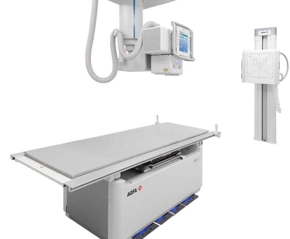
AGFA DR600
The DR600 digital radiography system from AGFA transforms the X-ray room into a versatile workspace through its automated workflow and intelligent ceiling system design. This innovative system integrates digital motor and spatial mapping technology, enabling the automatic movement of the tube and the detector to predefined positions, thus reducing patient waiting time. It can be used for examinations with wall-mounted detector configurations as well as radiography on a movable examination table. In addition, due to its ZeroForce technology, it facilitates the effortless relocation of the equipment without physical strain, while the touchscreen integrated into the tube allows easy selection of the X-ray protocol and expedites the overall procedure.
The system is equipped with two high-resolution wireless digital detectors, and in combination with the built-in automatic exposure control sensors, it automatically adjusts radiation parameters for optimal image quality at the lowest possible dose, adapting to the examinee’s body type.
The machine has additional filters and the option of anti-scatter grid to further improve diagnostic quality and reduce radiation dose.
AGFA DR100s
The portable X-ray machine is primarily used in the hospital wards where patients are immobile. The mobile system features digital motors that support easy movement of the equipment to patient beds. It is equipped with a wireless digital detector to provide immediate diagnostic information wherever it is located.

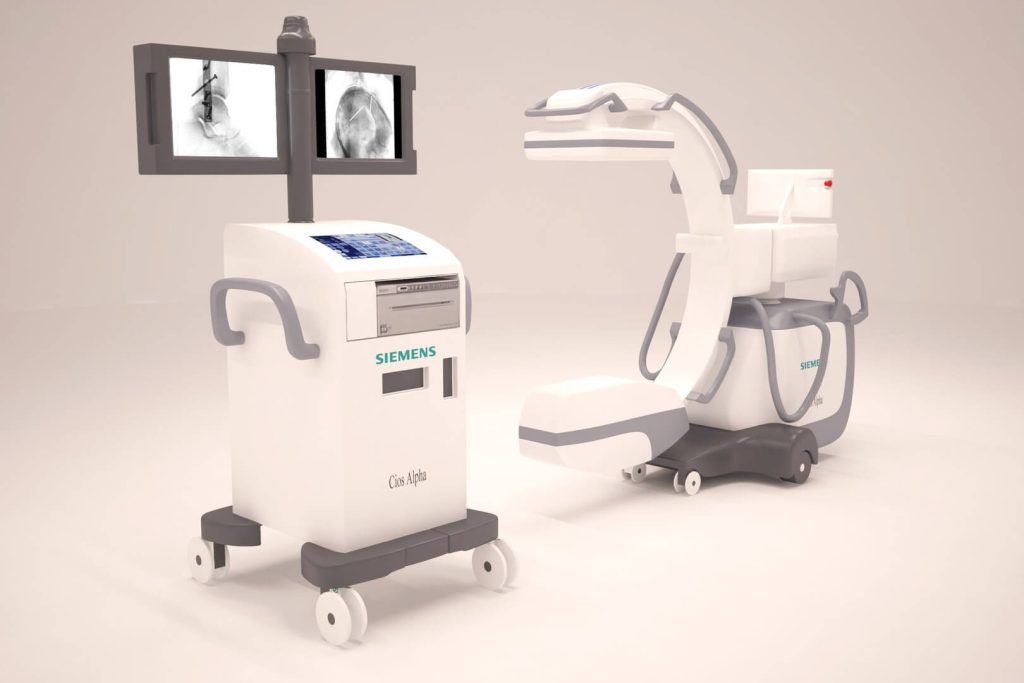
Siemens Cios Alpha mobile C-arm
In image-guided interventions, the trade-off between image quality and radiation dose is a common concern. Higher doses can pose health risks, while dose reduction often results in lower image quality. The Cios Alpha C-arm has been engineered to address this challenge effectively. Equipped with Retina technology, it ensures optimal image quality. It also employs CARE technologies to customise the radiation dose for each case, focusing on patient safety. Intelligent power management further enhances its capabilities, striking the perfect balance between image quality and dose.
The Cios Alpha offers a range of benefits. Alongside excellent image quality and minimal dose, it provides an easy-to-use platform with a streamlined workflow, reducing distractions during procedures. Its design concentrates on the patient rather than the equipment, saving time and enhancing efficiency.
The Cios Alpha also excels in data handling and connectivity, making the transfer of information seamless.
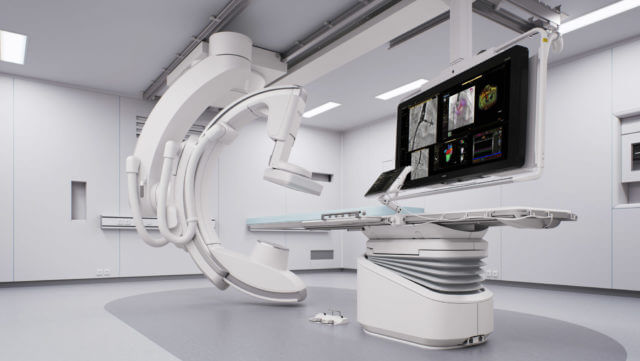
Angiography
The angiographic systems within the facility present the capacity for a broad range of interventional procedures with the highest possible accuracy, minimal radiation exposure, and the expedited duration, providing ease of operation for the physician and reduced risk to the patient. These systems encompass two of Philips’ most advanced and modern platforms, which come with the pioneering ClarityIQ technology that significantly reduces the radiation dose while maintaining the highest diagnostic image quality. Both systems are equipped with innovative software applications that enable qualitative and quantitative measurements for treatment evaluation such as 2D Perfusion for vascular and tissue perfusions, Aneurysm Flow for assessing blood flow dynamics before and after the treatment, and various other tools to assist in interventional procedure such as 3D Road mapping for real-time 3D navigation and the cone beam angiography technique for CT-type images designed to enhance visualisation of internal anatomical structures.
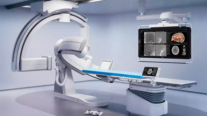
Azurion 7 B20/15
Our facility stands as the first centre in Cyprus to house the cutting-edge Biplane digital angiography system. This signifies a remarkable advancement in interventional radiology and interventional neuroradiology, offering the possibility for innovative procedures and treatments, with the highest possible safety for the patient. It features simultaneous imaging in two planes, offering top-quality image with a significantly reduced radiation dose.
Azurion 7 C20
This is a modern hybrid ceiling system developed by Philips, enabling the capability for both angiography and surgical procedures. The flexibility of the robotic arm guarantees maximum precision for physicians, all while minimising radiation exposure to patients.
Ultrasound
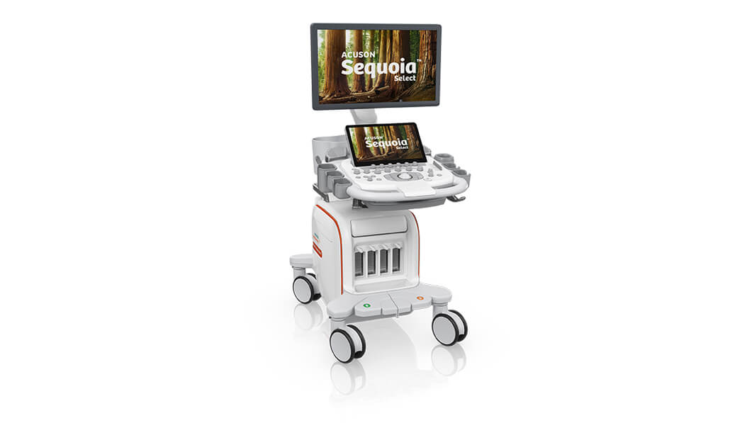
Siemens Acuson Sequoia / Juniper – Philips Sparq
The centre is equipped with modern portable ultrasound systems featuring advanced applications, offering physicians the flexibility to perform a wide range of diagnostic and interventional procedures. These include Siemens Acuson Sequoia and Juniper, and Philips Sparq. These systems have high-resolution transducers, which can operate in a wide range of frequencies and transducer to suit various anatomies and examinations. They incorporate innovative features, which enable high-resolution imaging tailored to each individual’s anatomy and body type. Advanced technologies like shear wave elastography and colour Doppler are also available. The Acuson Sequoia further features a navigation system with electromagnetic sensors that record the needle’s position, allowing for its orientation in a three-dimensional space to be derived from MRI or CT images, and thus ensuring precision in needle guidance.
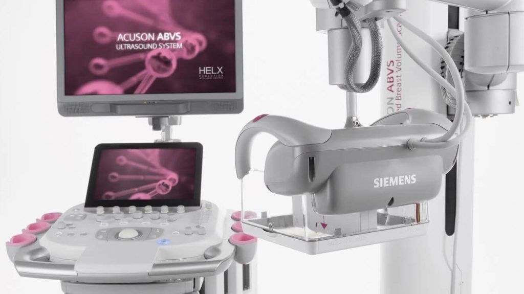
Siemens Automated Breast Volume System (ABVS) S2000
The department also has at its disposal the Siemens S2000 ABVS ultrasound system. This is an automated three-dimensional breast ultrasound and represents the most advanced and innovative ultrasound system technology for the prevention and diagnosis of breast cancer. The 3D imaging allows evaluation of the breast in the transverse, coronal and sagittal planes, with thin slice reconstruction. This enables the detection of very small lesions that may not be on a standard ultrasound, and provides precise localisation of lesions within the breast, which is invaluable information for surgical interventions.
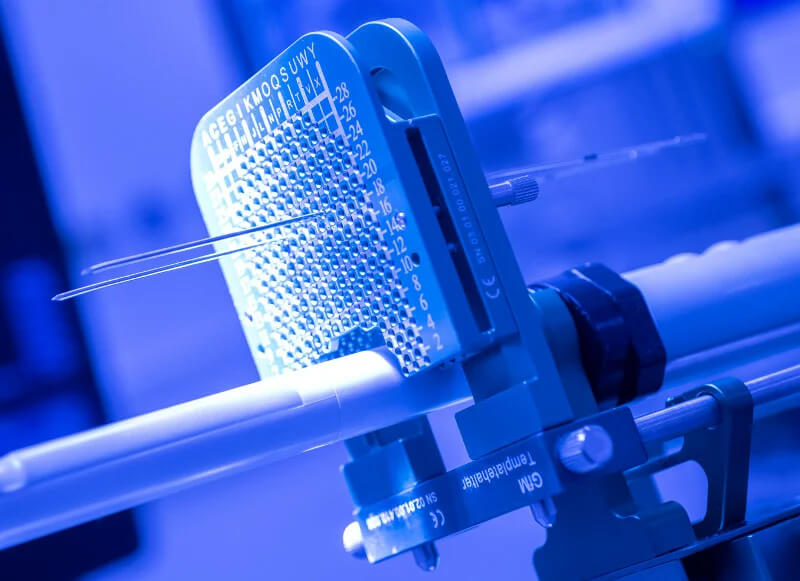
MEDCOM GMBH MobileUS
The department also houses the MEDCOM GMBH MobileUS ultrasound, with a dedicated stand for transperineal prostate biopsies. This system is equipped with specialised software enabling the fusion of ultrasound and MR images, allowing for detailed contouring of the anatomy of the prostate and the identification of potential lesions within the prostate, the planning of the biopsy procedure, and provides coordinates for accurate needle placement in the designated sampling areas.